
World's first field-installed, clinically-approved parallel transmit 7T MRI scanner resides at Auburn University
Published: Mar 26, 2024 8:20 AM
By Joe McAdory
Auburn University is now home to the world’s first field-installed, clinically-approved parallel transmit Terra.X 7 Tesla magnetic resonance imaging scanner — expanding the window for cutting-edge research and public use.
The $9 million Siemens MAGNETOM 7T, located in the Auburn University Samuel Ginn College of Engineering’s Neuroimaging Center, was certified by the Food and Drug Administration for use on clinical patients and it provides superior imaging compared to the college’s previous 7T research device. Additionally, with dedicated radiofrequency sodium imaging coils and parallel transmit technology, the scanner offers expanded imaging capabilities.
“Today, we can say the best MRI scanner in the world is sitting here in the Research Park in Auburn,” said Steve Taylor, senior vice president for research and economic development. “Impactful, multi-disciplinary research remains one of the foundations of our rich history as a university, and the door is now open to creative explorations into neuroscience that we, nor peer institutions, could previously pursue. I’m excited to watch researchers from across campus take advantage of this powerful resource.”
Predominant use of the new 7T will be for brain and knee imaging, with a clinical focus on better understanding epilepsy, multiple sclerosis and other disorders, said Thomas Denney, director of the Neuroimaging Center who also serves as the Bruce Donnellan and Family Professor in the Department of Electrical and Computer Engineering.
“The university is sitting on a gold mine in terms of data and capabilities,” Denney said. “This allows us to play in a sandbox that we couldn’t play in before. Our vision is to become a leading center for MRI research with emphasis on brain imaging, cardiovascular imaging, and orthopedic imaging. These areas represent an intersection between the needs of the MRI research community, particularly in 7T imaging, and existing expertise and strengths of Auburn University. In terms of biomedical research, this is one of those things that takes us to that next level.”
New research explorations already under way include, but are not limited to:
- Meredith Reid, assistant professor in electrical and computer engineering, who is exploring post-traumatic stress disorder biomarkers in senior adults via spectroscopy.
- Jennifer Robinson, professor in psychological sciences in the College of Liberal Arts, who is comparing brain connectivity between healthy populations with mental illness with a focus on the interaction between cognition and emotions.
- Adil Bashir, associate professor in electrical and computer engineering, who is studying muscle and brain energy production capacity on the cells and determining mitochondrial metabolic homeostasis by utilization of phosphorus spectroscopy.
- Gopikrishna Deshpande, professor in electrical and computer engineering, who is examining how artificial intelligence can be used to predict brain disorders and human/animal behavior using information from brain networks obtained from MRI.
- Doug Martin, director of the Scott-Ritchey Research Center in the College of Veterinary Medicine and professor of Anatomy, Physiology & Pharmacology, who is measuring the effect of gene therapy for Tay-Sachs disease in animal models using magnetic resonance imaging and spectroscopy.
“In addition to being able to provide opportunities for researchers like myself to study disorders and even brain connectivity at a new level, this new scanner also contributes to the fact that we can clinically assess people in different ways,” said Robinson, principal investigator at the Auburn University Cognitive & Affective Neuroscience Laboratory. “It has tremendous impacts for folks with epilepsy or Parkinson's where we can do imaging that we couldn't do before. That's a tremendous asset for the state of Alabama and especially for our area. Those things, both the research aspect and the clinical aspect, make it a pivotal resource.”
The Center’s previous 7T system, installed in 2012 and removed, was an investigational-only device, where subjects were scanned for research, not clinical, purposes.
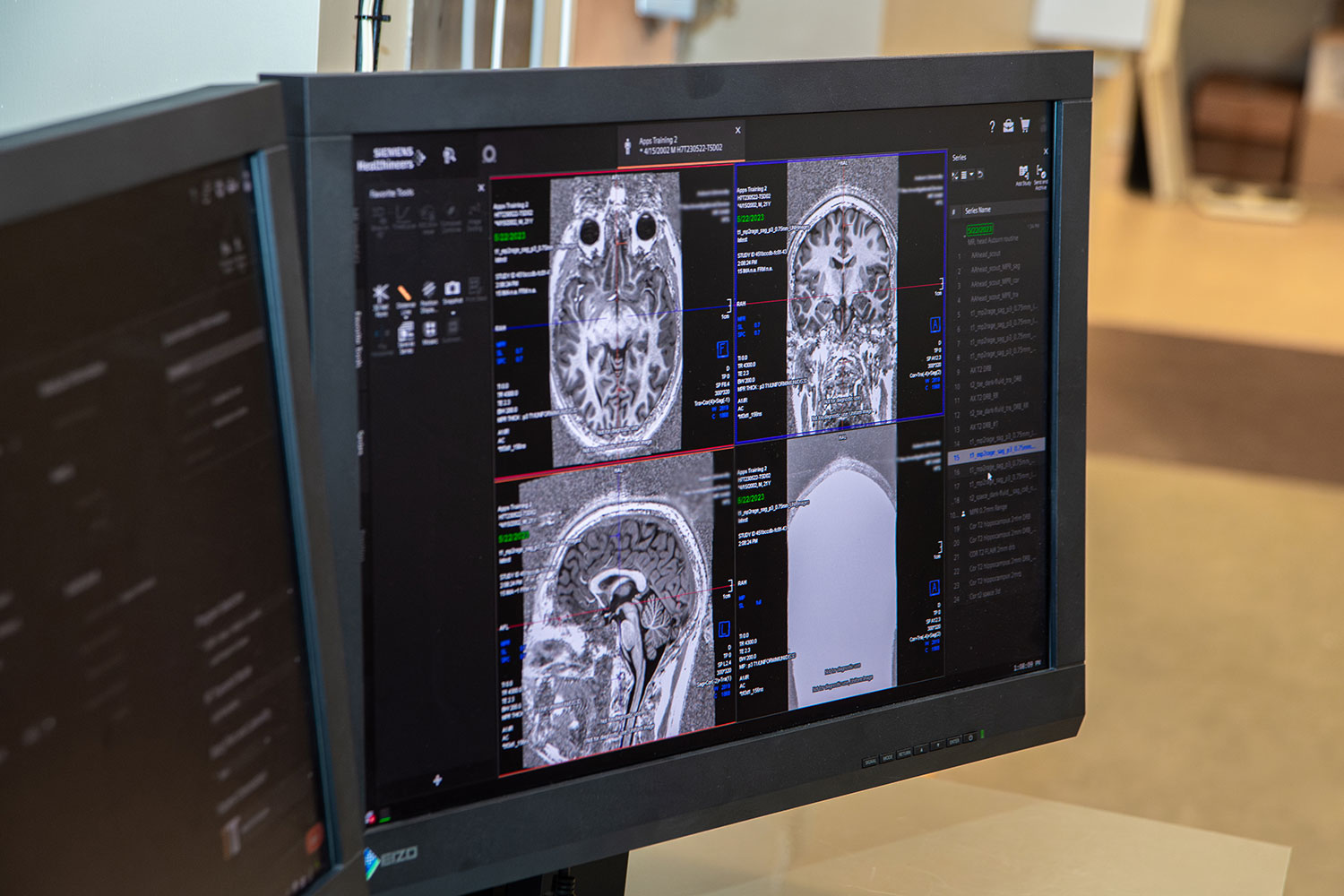
“There will be physicians who will want to send their patients here to be scanned on the new 7T,” Denney said. “People have MRI scans to answer clinical questions. If a question can be answered with a 3T, then that respective hospital will more than likely have that equipment. But if the 3T does not answer the question — as our 7T provides deeper and clearer images — then those patients would benefit from our device.”
A stark difference between the old and new 7T scanners involve parallel transmit technology and sodium imaging capability.
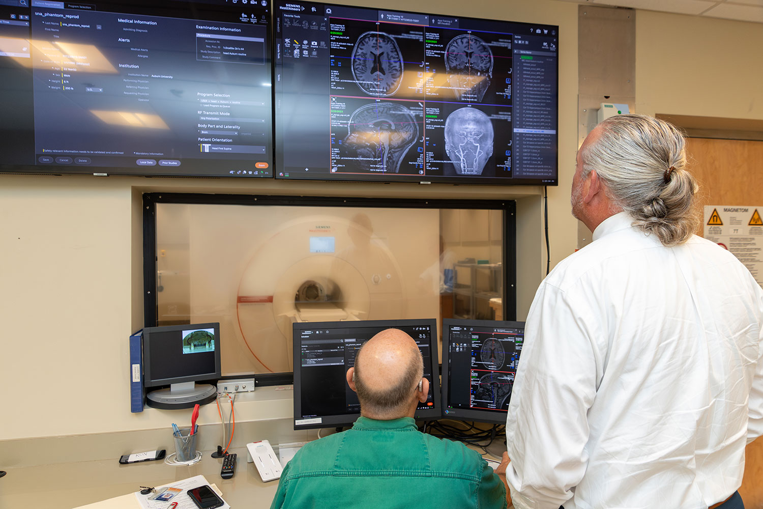
“With our old 7T system, if we did an MRI scan of the brain in certain regions, you wouldn’t receive uniform coverage of the brain,” Denney said. “That’s because we had only one transmit channel, like a flashlight inside a large room with only one beam. By comparison, our new Terra.X 7T provides us with eight beams of light that illuminates the brain and provides a more uniform image.
“Sodium imaging technology is also important because it can measure how much sodium is in your brain regionally. This allows us to identify abnormalities and study electrical signaling in the brain,” Denney added.
A gift from Auburn Engineering alumnus Thomas Walter, ’55 engineering physics, and his wife, Jean, ’57 education, paved the way for the addition of the new machine to the 45,000-square foot building, and, in turn, the university named the facility in his honor as the Thomas Walter MRI Research Building.
“We are certainly honored and humbled to have alumni and friends like Tom and Jean Walter,” said Mario Eden, dean of the Samuel Ginn College of Engineering. “If it wasn’t for them, we wouldn’t have the 7T, and we are indebted to them for their dedication and commitment to Auburn. Through this cutting-edge technology, we are fortunate to have the opportunity to show how we can utilize their generosity for the betterment of mankind through impactful research.”
Media Contact: , jem0040@auburn.edu, 334.844.3447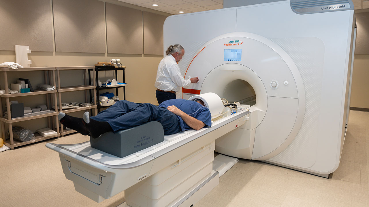
With dedicated radiofrequency sodium imaging coils and parallel transmit technology, the Terra.X 7 Tesla scanner offers expanded imaging capabilities.